The Role of Advanced Diagnostic Equipment in Identifying Eye Disorders
In the world of ophthalmology, the utilization of innovative analysis tools has transformed the very early identification and management of different eye disorders. From discovering subtle modifications in the optic nerve to keeping track of the progression of retinal illness, these innovations play an essential function in improving the precision and performance of diagnosing ocular problems. As the demand for specific and prompt medical diagnoses continues to expand, the assimilation of sophisticated devices like optical coherence tomography and visual field testing has actually come to be crucial in the world of eye treatment. The elaborate interaction in between innovation and sensory practices not just sheds light on elaborate pathologies but additionally opens up doors to tailored therapy techniques.
Relevance of Early Diagnosis
Early diagnosis plays a critical role in the effective management and treatment of eye problems. By spotting eye problems at a very early stage, healthcare service providers can offer proper treatment strategies tailored to the certain problem, inevitably leading to better outcomes for people.

Innovation for Identifying Glaucoma
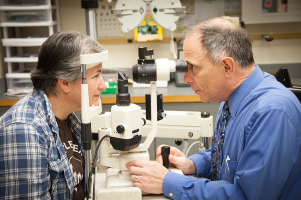
Sophisticated diagnostic technologies play a vital function in the early discovery and surveillance of glaucoma, a leading source of permanent loss of sight worldwide. One such modern technology is optical coherence tomography (OCT), which supplies comprehensive cross-sectional pictures of the retina, enabling the measurement of retinal nerve fiber layer thickness. This measurement is vital in assessing damages brought on by glaucoma. Another sophisticated device is aesthetic area testing, which maps the level of sensitivity of a client's aesthetic field, assisting to identify any areas of vision loss attribute of glaucoma. Furthermore, tonometry is used to gauge intraocular pressure, a major danger aspect for glaucoma. This examination is critical as elevated intraocular pressure can bring about optic nerve damage. Newer innovations like the usage of fabricated knowledge algorithms in evaluating imaging information are showing encouraging results in the very early detection of glaucoma. These sophisticated analysis tools make it possible for eye doctors to diagnose glaucoma in its early phases, permitting for timely intervention and much better administration of the illness to stop vision loss.
Function of Optical Comprehensibility Tomography

OCT's capacity to evaluate retinal nerve fiber layer density permits accurate and unbiased measurements, assisting in the very early discovery of glaucoma even before visual field problems become obvious. OCT technology allows longitudinal surveillance of structural adjustments over time, promoting customized therapy strategies and timely interventions to aid protect patients' vision. The non-invasive nature of OCT imaging also makes it a favored selection for keeping track of glaucoma progression, as it can be duplicated regularly without creating discomfort to the individual. On the whole, OCT plays an essential duty in improving the diagnostic accuracy and monitoring of glaucoma, ultimately adding to far better results for people in danger of vision loss.
Enhancing Diagnosis With Visual Field Screening
A vital component in comprehensive ophthalmic evaluations, aesthetic area testing plays a critical duty in enhancing the diagnostic procedure for numerous eye conditions. By assessing the complete level of a client's aesthetic field, this test offers crucial information about the useful honesty of the whole aesthetic pathway, from the retina to the aesthetic cortex.
Visual field screening is especially useful in the medical diagnosis and management of conditions such as glaucoma, optic nerve conditions, and different neurological conditions that can affect vision. With measurable dimensions of outer and central vision, clinicians can find subtle modifications that may indicate the presence or progression of these disorders, even prior to visible signs and symptoms happen.
Furthermore, visual field testing enables the tracking of therapy efficiency, helping ophthalmologists customize therapeutic interventions to private clients. eyecare near me. By tracking changes in visual area efficiency gradually, doctor can make educated choices about adjusting medications, advising medical interventions, or implementing other suitable steps to protect or improve a client's visual function
Managing Macular Deterioration

Final Thought
In verdict, progressed diagnostic devices play an important duty in recognizing eye disorders early on. Technologies such as Optical Coherence Tomography and aesthetic area testing have considerably enhanced the precision and effectiveness of detecting conditions like glaucoma and macular deterioration.